Efficiently diagnose with awareness of patient conditions. Finding microscopic nodules provides a variety of information, including basic, number of nodules, size and status, and RADS category. Findings that are likely to develop into lung cancer can also be checked in advance, reducing working time and allowing efficient reading depending on the case. Microscopic Nodule Detection: Providing Comprehensive Information on Number of Nodules, Size, Status, and RADS Category Early Detection of Potential Lung Cancer Development Enables Time-efficient Scans Reading.
Efficiently diagnose with awareness of patient conditions. Finding microscopic nodules provides a variety of information, including basic, number of nodules, size and status, and RADS category. Findings that are likely to develop into lung cancer can also be checked in advance, reducing working time and allowing efficient reading depending on the case. Microscopic Nodule Detection: Providing Comprehensive Information on Number of Nodules, Size, Status, and RADS Category Early Detection of Potential Lung Cancer Development Enables Time-efficient Scans Reading.
Automate Tedious manual Tasks to Save Time.
Time Saving
Sensitivity
False positivity rate
Reduce Workload
https://dailybulletin.rsna.org/db22/index.cfm?pg=22fri07
Lancaster HL, Zheng S, Aleshina OO, Yu D, Yu Chernina V, Heuvelmans MA, de Bock GH, Dorrius MD, Willem Gratama J, Morozov SP, Gombolevskiy VA, Silva M, Yi J, Oudkerk M. Outstanding negative prediction performance of solid pulmonary nodule volume AI for ultra-LDCT baseline lung cancer screening risk stratification. Lung Cancer. 2022 Jan 6;165:133-140
BoneView represents Gleamers first application in the clinical AI domain. It has evolved into a global standard for the interpretation of X-ray images in cases of bone injuries, and is appreciated for its scientific excellence.
This AI application efficiently identifies fractures, effusions, dislocations, and bone lesions.
Recognized for its scientific rigor through publications in top-tier, peer- reviewed journals, its clinical study was honored with the prestigious Alexander Margulis Award 2022 for scientific excellence.
Patient-centred workflow Enables same day diagnostics for patients
Reducing unnecessary MRI examinations Only offering MRIs to the patients who will benefit the most
Increased Standardisation
Unified pathway for patients with knee osteoarthritis
At the core of BrainScan CT are sophisticated AI algorithms, adept at scanning, identifying, and pinpointing potential pathological changes within the brain.
The comprehensive analysis results are provided in dual formats: visually engaging infographics and detailed structured text, all returned to the originating PACS server. This dual-format approach ensures thoroughness and clarity in the diagnostic process.
Streamlining the breast imaging workflow – saves time on operational tasks and communications.
Standardizes procedures in mammography and breast ultrasound, compatible across all mammography vendors – enhances overall efficiency.
Automates the creation of audit-ready documentation in mammography – reduces costs associated with regulatory compliance and reporting.
Decreases the rate of misdiagnosis through real-time quality assurance in mammography and by stratifying high-risk patients.
Enhances quality performance using B-Rayz's advanced technology-driven learning platform.
Provides SUVR quantification report of various PET tracers observed in PET images.
Reduce Patient Recalls
Acquiring the appropriate image during the initial examination accelerates and optimizes the diagnostic process, as the radiologist possesses the necessary information to complete their assessment without the need to recall the patient for additional imaging.
Radiographers Decision Support
Smart Protocol offers real-time decision support to radiographers, helping them make informed decisions about additional imaging or other procedural adjustments limiting unnecessary interruption of radiologists throughout the imaging process.
Scan Time / Protocol Optimization
Smart Protocol optimizes scan time by adding only necessary sequences when critical conditions are detected. These dynamic adjustments ensure comprehensive diagnostics while improving efficiency
Improved Patient Journey
Tailored protocols based on individual patient data lead to personalized and more effective diagnostic patient journeys
Patient Satisfaction
Minimizing the need for multiple visits for diagnostics improves patient convenience and satisfaction. Acquiring the right number of sequences helps reduce patient discomfort due to lengthy exams.
Automatically segments the brain in MRI, providing quantitative measures of brain volume and white matter hyperintensities.
Customized analysis reports for users.
LCS Plus is a trusted solution in global lung cancer screening projects.


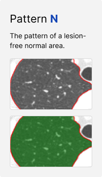
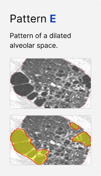
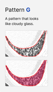
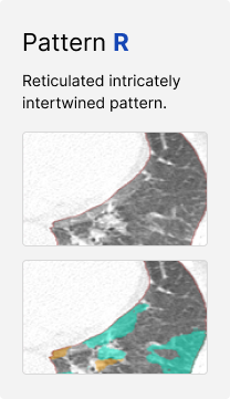
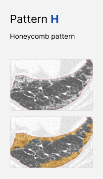
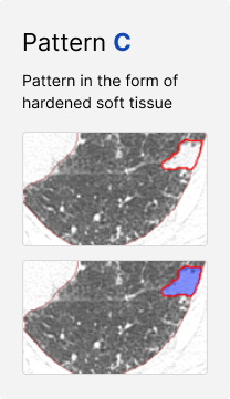
The use of advanced algorithms enables the automated analysis of the locations and distribution of the six recognized patterns associated with ILD.
By standardizing the analyses and minimizing variations in interpretation, this solution ensures accurate and reliable results.
The product improves inter- and intra-operator consistency in reporting and grading by identifying and classifying the following organisms:
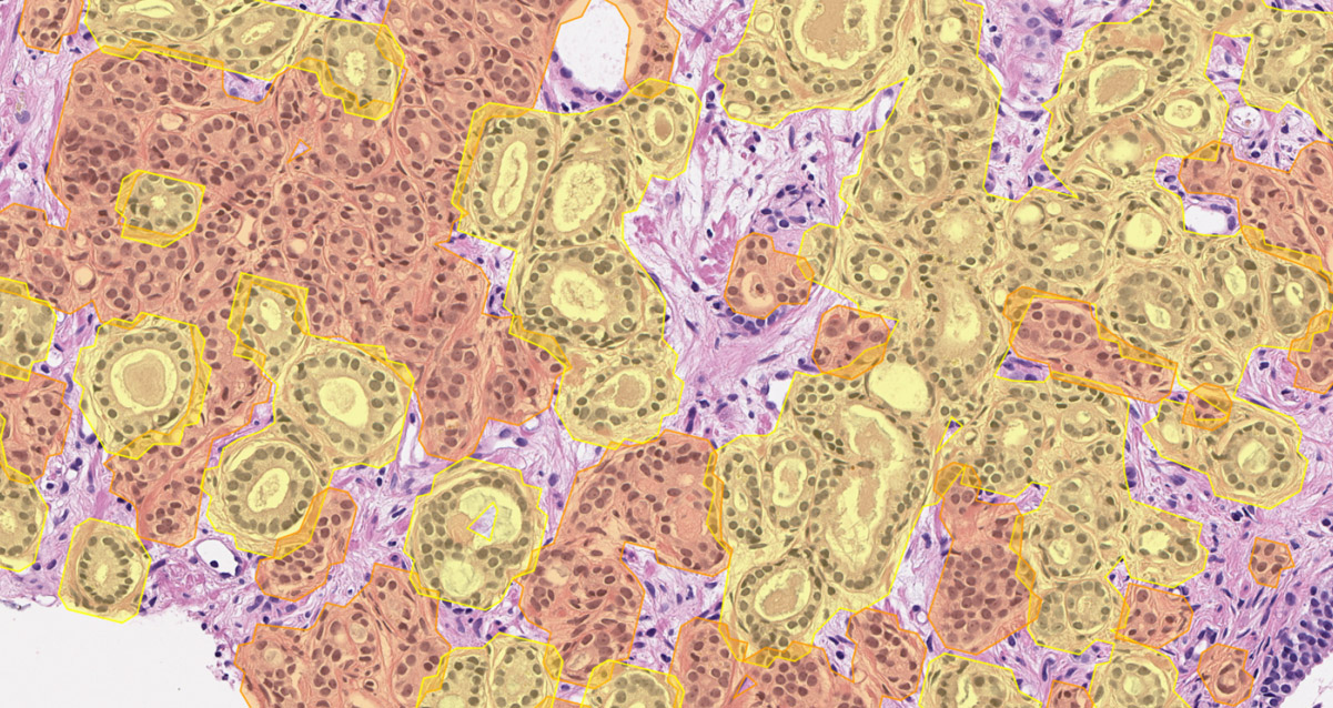
Pre-filled pathology reports are generated once slides are analyzed using DeepDx® Prostate. Clinically significant information included in the pathology report includes:
• Gleason scores
• Relative proportions of each Gleason pattern
• Percentage involvement of cancer in tissue
Quantitative metrics such as tumor-to-tissue ratios and Gleason pattern ratios are recalculated in real-time as annotations are made.
Sample images representative of the cancer found in a slide.
ICC
Consistency
Suh YJ, Kim C, Lee JG, Oh H, Kang H, Kim YH, Yang DH. Fully automatic coronary calcium scoring in non-ECG-gated low-dose chest CT: comparison with ECG-gated cardiac CT. Eur Radiol. 2023 eb;33(2):1254-1265. doi: 10.1007/s00330-022-09117-3. Epub 2022 Sep 13.
Abnormal findings detecting in chest X-ray images
Digital hematology manual differential assistance for diagnosis
Enhancing Diagnostics through Intelligent Image Analysis
AI-based analysis to support the diagnosis of interstitial lung diseases