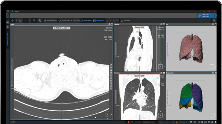
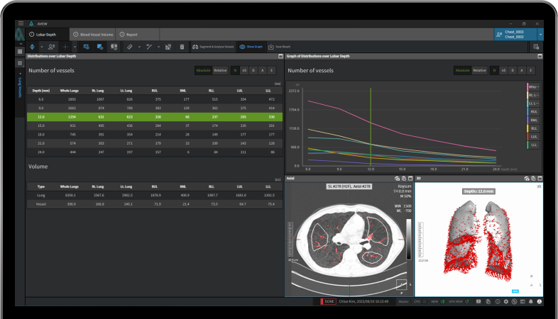
Automated Lungs, Lung Lobes, and Pulmonary Vascular Airways Segmentation.
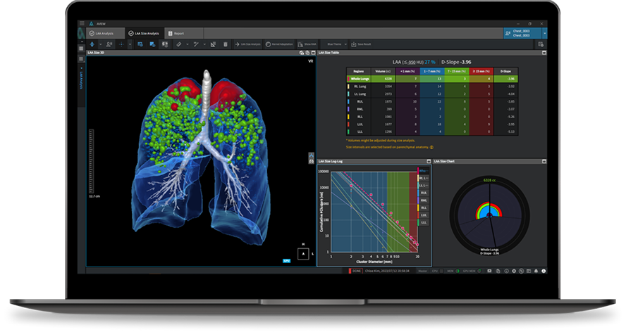
Utilize 3D Emphysema Visualization to Observe Its Distribution and Track Disease Progression. Enhance Understanding of Lungs and Lung Lobes with Visualized Content, Including Images and Graphs.


About 4,000 Analysis Results exported in CSV.
Export Quantitative Analysis Metrics for Research on Pulmonary Function. Utilize Detailed Analysis Reports in PDF and DICOM Formats for Clinical and Research Purposes.
Pulmonary emphysema
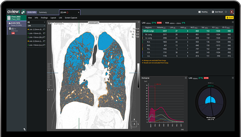
LAA
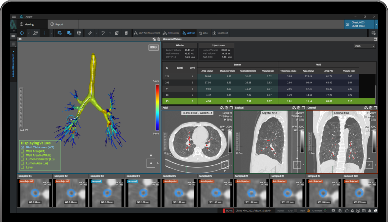
Airway
AWT-Pi10
Heejun Park, MS, Jaeyoun Yi, PhD, Donghoon Yu MS, Hyungi Seo MS, Jongha Park, MS, Jihye Yun, PhD, Namkug Kim, PhD, Sang Min Lee A, MD, Sang Min Lee B, MD, Joon Beom Seo, MD, PhD, "Fully Automated Workflow for Advanced Quantitative Analysis on Multi-Volume Chest CT of Patients with Chronic Obstructive Pulmonary Disease using Deep Convolutional Neural Net and Conventional Image Processing" RSNA2018, Chicago I
Augmenting Mammography with Artificial Intelligence
Digital ova and parasite identification assistance for diagnosis
Diagnosis of neurodegenerative diseases
Smart Ultrasound software providing early and accurate breast cancer diagnosis.